Lutein: The Macular Carotenoid Antioxidant
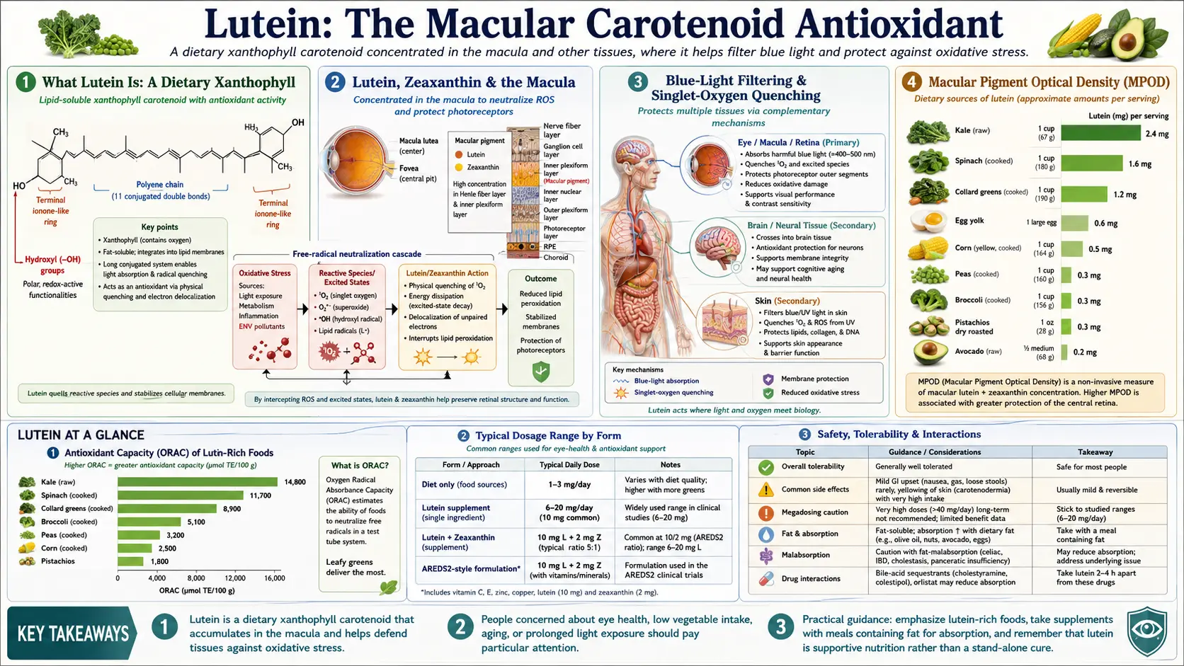
Lutein is a yellow xanthophyll carotenoid that the human body cannot synthesize and must obtain from food. It is concentrated — together with its stereoisomer partner zeaxanthin — in the macula of the retina, where the two pigments form the macular pigment that filters damaging blue light and quenches singlet oxygen. Lutein is also the dominant carotenoid in human brain tissue and in breast milk, and it accumulates in skin. The strongest clinical evidence comes from the AREDS2 trial (JAMA, 2013), in which lutein plus zeaxanthin reduced progression to advanced age-related macular degeneration and safely replaced beta-carotene in the AREDS formula.
Interactive Visualization Vision & the Retina — focus light and watch rods fire Follow the phototransduction cascade as rods and cones react, then dim the lights and watch the eye adapt. Launch →
Table of Contents
- What Lutein Is: A Dietary Xanthophyll
- Lutein, Zeaxanthin & the Macula
- Blue-Light Filtering & Singlet-Oxygen Quenching
- Macular Pigment Optical Density (MPOD)
- Age-Related Macular Degeneration & AREDS2
- Cataract Risk
- Blue Light, Screens & Visual Performance
- Brain & Cognition
- Skin & Photoprotection
- Dietary Sources & Bioavailability
- Forms, Dosage & Absorption
- Safety and Cautions
- Key Research Papers
- Connections
- Featured Videos
What Lutein Is: A Dietary Xanthophyll
Lutein (pronounced "LOO-teen," from the Latin luteus, meaning yellow) is one of more than 600 carotenoids found in nature and one of roughly two dozen that circulate in human blood. Carotenoids divide into two families: the carotenes, which are pure hydrocarbons (for example beta-carotene and lycopene), and the xanthophylls, which carry oxygen atoms on their ring structures. Lutein is a xanthophyll: its molecular formula is C₄₀H₅₆O₂, with a hydroxyl group on each of its two end rings. Those oxygen-bearing rings make lutein more polar than the carotenes and steer how it orients itself inside cell membranes.
Crucially, humans — like all animals — cannot make lutein. Plants, algae, and some microbes synthesize it; we acquire it entirely through diet, concentrate it in specific tissues, and have evolved binding proteins that selectively capture it. Lutein is what gives marigold petals, egg yolks, and corn their yellow color, and it contributes to the green of leafy vegetables, where it is masked by chlorophyll.
Lutein almost always travels with zeaxanthin, a closely related xanthophyll that differs only in the position of one double bond in one of the end rings. The two are so similar that nutrition science and the major clinical trials treat them as a pair (“L/Z”). A third relevant molecule, meso-zeaxanthin, is not generally found in food at all — the body manufactures it in the retina by converting lutein, which underscores how central lutein is to the eye's chemistry. Because zeaxanthin is lutein's inseparable partner, the two are best understood together; see Zeaxanthin.
Lutein, Zeaxanthin & the Macula
The macula is the small, roughly 5 mm region at the center of the retina responsible for sharp central vision, color vision, and fine detail. At its very center lies the fovea, where cone photoreceptors are packed most densely. The macula is yellow — the Latin name macula lutea literally means "yellow spot" — and that yellow color comes entirely from accumulated lutein, zeaxanthin, and meso-zeaxanthin. Collectively these three are the only carotenoids found in the macula, even though dozens circulate in the bloodstream. The retina is therefore extraordinarily selective: it filters out lycopene, beta-carotene, and every other carotenoid and admits only the xanthophyll trio.
The spatial distribution is precise. Meso-zeaxanthin and zeaxanthin dominate at the very center of the fovea; lutein predominates in the surrounding parafoveal and peripheral retina. This layered arrangement is thought to optimize protection across the macula because the different isomers absorb blue light slightly differently and orient differently within the photoreceptor membranes.
Two retinal binding proteins explain the selectivity. StARD3 (also called MLN64) binds lutein, while GSTP1 (a pi-class glutathione S-transferase) binds zeaxanthin and meso-zeaxanthin. These dedicated carriers escort the xanthophylls into the photoreceptor and pigment-epithelium layers and hold them in place, which is why dietary intake translates into measurable macular accumulation over weeks to months.
This concentration is not incidental. The macula sits directly in the path of focused light, the photoreceptors there have the highest metabolic rate and oxygen consumption of any cells in the body, and the outer-segment membranes are packed with easily oxidized polyunsaturated fatty acids such as DHA. That combination — intense light, high oxygen, abundant oxidizable lipid — makes the macula one of the most oxidatively stressed tissues anywhere. The xanthophylls are its built-in optical and chemical shield.
Blue-Light Filtering & Singlet-Oxygen Quenching
Lutein protects the retina through two complementary mechanisms — one optical, one chemical.
1. Blue-light filtering (the optical shield). Lutein and zeaxanthin absorb strongly in the blue region of the visible spectrum, with peak absorption around 445–460 nm. Short-wavelength blue light carries the most energy per photon of any visible light and is the most damaging to the retina; it also scatters most inside the eye, degrading contrast. By sitting in a layer in front of the photoreceptors, the macular pigment acts like internal sunglasses — absorbing a substantial fraction of incoming blue light before it can reach and damage the sensitive outer segments and the underlying retinal pigment epithelium. A useful side effect is reduced glare and improved contrast sensitivity, because the scattered blue haze is filtered out.
2. Singlet-oxygen quenching and antioxidant action (the chemical shield). When light strikes the retina, it generates reactive oxygen species, especially singlet oxygen — an excited, highly reactive form of O₂. Carotenoids are among the most efficient singlet-oxygen quenchers known: their long chain of conjugated double bonds absorbs the excess energy from singlet oxygen and dissipates it harmlessly as heat, regenerating the intact carotenoid. Lutein and zeaxanthin also directly scavenge peroxyl and other free radicals, interrupting the chain reaction of lipid peroxidation in the photoreceptor membranes. Because their hydroxylated rings let them span and orient within the lipid bilayer, they are positioned exactly where membrane lipids are most vulnerable.
This dual action makes lutein conceptually similar to other lipid-phase carotenoid antioxidants such as astaxanthin, which is also studied for eye and skin photoprotection. Where lutein is unique is its selective, protein-mediated deposition in the macula and brain.
Macular Pigment Optical Density (MPOD)
The amount of lutein and zeaxanthin actually deposited in the macula can be measured non-invasively and is expressed as macular pigment optical density (MPOD) — a number that quantifies how much blue light the pigment layer absorbs. MPOD is measured by techniques such as heterochromatic flicker photometry, autofluorescence imaging, and resonance Raman spectroscopy. It functions as a biomarker: higher MPOD reflects more protective pigment in place.
MPOD varies widely between individuals and is influenced by dietary intake, blood xanthophyll levels, age, smoking, body fat (carotenoids are sequestered in adipose tissue), and genetics of the binding and transport proteins. Importantly, MPOD is responsive to diet: supplementation trials consistently show that taking lutein and zeaxanthin raises serum levels within days and increases MPOD over roughly 8–24 weeks. This dose–response relationship — eat more, measure more in the eye — is one of the clearest in nutritional science and is the mechanistic backbone for the clinical eye-health trials.
Low MPOD has been associated with greater risk of age-related macular degeneration, while higher MPOD tracks with better visual performance metrics including contrast sensitivity, glare recovery, and reduced photostress recovery time.
Age-Related Macular Degeneration & AREDS2
Age-related macular degeneration (AMD) is the leading cause of irreversible central vision loss in older adults in the developed world. Because the macular pigment is composed of lutein and zeaxanthin, and because oxidative damage and blue-light exposure are central to AMD pathology, the xanthophylls became an obvious target for prevention research.
The AREDS and AREDS2 trials
The original Age-Related Eye Disease Study (AREDS, 2001) showed that a high-dose antioxidant and zinc formula (vitamin C, vitamin E, beta-carotene, zinc, copper) reduced the risk of progression to advanced AMD by about 25% in people at high risk. But the beta-carotene component was problematic: separate trials had shown that high-dose beta-carotene significantly increased lung-cancer risk in current and former smokers.
AREDS2 (Age-Related Eye Disease Study 2; Chew et al., JAMA, 2013) was designed to fix this. More than 4,000 participants at high risk of advanced AMD were randomized in a factorial design that tested adding lutein (10 mg) plus zeaxanthin (2 mg) and/or omega-3 fatty acids, and that tested removing or lowering beta-carotene. Key findings:
- Adding lutein + zeaxanthin to the formula provided protection against progression to advanced AMD comparable to beta-carotene — without beta-carotene's lung-cancer signal.
- In the direct comparison, substituting lutein + zeaxanthin for beta-carotene was associated with a roughly 18% further reduction in progression to advanced AMD.
- The benefit of lutein + zeaxanthin was greatest in participants with the lowest dietary intake of these carotenoids at baseline.
- Omega-3 fatty acids (DHA + EPA) showed no significant additional benefit on AMD progression in this trial.
The practical result was a rewritten standard of care: the modern AREDS2 formula replaces beta-carotene with 10 mg lutein and 2 mg zeaxanthin and is recommended for people with intermediate AMD or advanced AMD in one eye. It is one of the few supplement interventions with large, double-blind, randomized backing for slowing a sight-threatening disease. Note the important caveat: AREDS2 demonstrated slowed progression in people already at high risk; it did not establish that the formula prevents AMD from developing in low-risk eyes.
Cataract Risk
Lutein and zeaxanthin are the only carotenoids detected in the human lens, where the same blue-light-filtering and antioxidant properties that protect the retina are thought to protect lens proteins (crystallins) from photo-oxidative damage that drives cataract formation. Several large prospective cohorts — including the Nurses' Health Study and the Health Professionals Follow-Up Study — reported that people with the highest dietary intake of lutein and zeaxanthin had a modestly lower risk of needing cataract extraction, on the order of 20% for the highest versus lowest intake groups.
Randomized-trial evidence is weaker and mixed. The AREDS2 trial found no overall statistically significant effect of lutein + zeaxanthin on cataract progression in the full cohort, though a benefit emerged in the subgroup with the lowest baseline dietary intake. The reasonable summary is that lutein-rich diets are associated with lower cataract risk in observational data, the mechanism is plausible, but supplements have not been proven to prevent cataract in well-nourished populations.
Blue Light, Screens & Visual Performance
Modern life floods the eyes with artificial blue light from LED screens, smartphones, and energy-efficient lighting. Because the macular pigment's defining property is blue-light absorption, lutein and zeaxanthin have been studied for visual comfort and performance in heavy screen users.
The most-cited work is the Lutein, Zeaxanthin, and Visual Function (LZVF) study by Stringham and colleagues, in which healthy young adults who took lutein (10 mg) plus zeaxanthin (2 mg) for six months raised their MPOD and showed measurable improvements in contrast sensitivity, photostress recovery (how quickly vision returns after a bright flash), disability glare, and even self-reported reductions in eye strain, fatigue, and headache during prolonged computer work. Other randomized trials in screen-heavy populations have reported similar improvements in visual fatigue symptoms and glare tolerance.
This is a different claim from disease prevention: rather than slowing AMD, here lutein and zeaxanthin appear to improve everyday visual performance and comfort by increasing the pigment that filters glare-producing blue light and speeds recovery from bright-light exposure. The evidence is promising and mechanistically coherent, though trials are smaller and shorter than the AMD literature.
Brain & Cognition
One of the most striking facts about lutein is that the macula is not its only privileged tissue: lutein is the dominant carotenoid in the human brain. Analyses of brain tissue across the lifespan — from infants to centenarians — consistently find that lutein accounts for the largest share of total brain carotenoids, typically around half, far out of proportion to its share in the bloodstream. The brain, like the retina, selectively concentrates lutein.
In infants this is especially notable. Lutein is a major carotenoid in breast milk and in the developing infant brain, where it is preferentially deposited in regions involved in memory and learning. This has driven interest in lutein as a nutrient for neurodevelopment, and lutein is now added to some infant formulas.
In adults and older people, MPOD (the eye measurement) serves as a convenient proxy for brain lutein status, because retinal and brain levels correlate. Cross-sectional and longitudinal studies — including work from the Georgia Centenarian Study and several supplementation trials — have linked higher lutein status and higher MPOD to better performance on measures of memory, processing speed, and executive function, and small randomized trials of lutein (with or without zeaxanthin) have reported improvements in cognitive measures in older adults. The proposed mechanisms include lutein's antioxidant and anti-inflammatory action in neural membranes and a role in the efficiency of neuronal membranes rich in DHA. The evidence is suggestive rather than definitive, but the brain's deliberate accumulation of lutein strongly implies a functional role.
Skin & Photoprotection
Lutein and zeaxanthin also accumulate in skin, where they contribute to the body's antioxidant defenses against ultraviolet and visible (including blue) light. By quenching singlet oxygen and free radicals generated by light exposure, dietary and topical carotenoids help limit photo-oxidative damage, lipid peroxidation, and the inflammation that drives photoaging.
Controlled studies, including work combining oral and topical lutein and zeaxanthin, have reported improvements in skin hydration, elasticity, surface lipids, and a reduction in lipid peroxidation, along with a measurable increase in skin carotenoid content. The effect size is modest, and lutein is an adjunct to sun protection rather than a replacement, but the same blue-light and singlet-oxygen chemistry that protects the eye operates in the skin.
Dietary Sources & Bioavailability
Because the body cannot synthesize lutein, diet is the only source. The richest foods are dark leafy greens, in which lutein and zeaxanthin are abundant but masked by chlorophyll.
- Cooked kale — among the most concentrated dietary sources, supplying on the order of 15–25 mg of lutein + zeaxanthin per cooked cup.
- Cooked spinach — another top source, roughly 12–20 mg per cooked cup; far more concentrated cooked (and wilted) than raw because the volume collapses.
- Other greens — collard greens, turnip greens, Swiss chard, romaine lettuce, parsley, and broccoli.
- Yellow and orange produce — corn (sweet corn is notably high in zeaxanthin), yellow peppers, and orange-fleshed squash.
- Egg yolk — lower in absolute lutein content than greens, but with markedly higher bioavailability (see below).
Bioavailability: why egg yolk punches above its weight
The amount of lutein in a food is only half the story; absorption matters as much. Lutein is fat-soluble, so it is absorbed far better when eaten with dietary fat. In leafy greens the lutein is bound up inside the plant's cell walls and chloroplast membranes, which limits how much is released and absorbed — cooking and chopping help break these structures down, and adding oil or another fat substantially increases uptake.
In egg yolk, by contrast, the lutein is already dissolved in the yolk's lipid matrix (phospholipids and cholesterol), delivered essentially pre-emulsified. Multiple controlled feeding studies have shown that the lutein in eggs is among the most bioavailable of any food source — gram for gram of lutein, eggs raise serum lutein and MPOD more efficiently than supplements or vegetables. Adding one or two eggs per day to the diet reliably increases blood lutein and zeaxanthin and, over weeks, macular pigment. This is why eggs feature so prominently in lutein nutrition despite their modest absolute content. (Older concerns about egg cholesterol have been substantially revised for most people; see the Eggs page.)
Forms, Dosage & Absorption
Supplemental lutein is most often extracted from marigold flowers (Tagetes erecta), whose petals are exceptionally rich in lutein. It is sold in two chemical forms:
- Lutein esters — the form as it naturally occurs in marigold, with fatty acids attached to the hydroxyl groups. Esters must be cleaved by pancreatic enzymes in the gut before the free lutein can be absorbed, so they are best taken with a fat-containing meal. They are stable and cost-effective, and on a per-milligram-of-lutein basis (after accounting for the weight of the attached fatty acids) they deliver lutein comparably to the free form when taken with food.
- Free (non-esterified) lutein — the form found in the bloodstream and in green vegetables. It does not require enzymatic cleavage. Labels for free lutein products report the actual lutein content directly; ester products should state the lutein equivalent.
One practical point on labeling: an ester product may list a higher total weight than its lutein equivalent because the fatty-acid portion adds mass, so compare the stated lutein (or "free lutein equivalent") rather than total ester weight.
Typical dosing:
- AREDS2 eye-health formula — 10 mg lutein + 2 mg zeaxanthin per day (the validated clinical dose for intermediate or advanced AMD).
- General eye and visual-performance support — 6–20 mg lutein per day, often with 2–4 mg zeaxanthin.
- Dietary baseline — population intake studies suggest most adults consume only 1–3 mg/day, well below the amounts associated with benefit, which is the rationale for emphasizing greens, eggs, or supplements.
Always take lutein with a meal containing fat to maximize absorption, and expect MPOD to rise gradually over 2–6 months rather than immediately. Lutein and zeaxanthin share absorption pathways with other carotenoids (notably beta-carotene), so very high doses of one carotenoid can modestly compete with the others for uptake.
Safety and Cautions
Lutein has an excellent safety record. It is a normal dietary constituent consumed by humans for our entire evolutionary history, and the U.S. and European food-safety authorities have affirmed its safety as a supplement and food additive. No tolerable upper intake level has been deemed necessary, and a Joint FAO/WHO expert committee established an acceptable daily intake of up to 2 mg per kilogram of body weight per day for lutein from marigold, far above typical supplement doses.
- Carotenodermia — very high, prolonged intake of carotenoids can cause a harmless yellow-orange tint to the skin (most visible on the palms and soles). It is benign and reverses when intake is reduced; it should not be confused with jaundice, which yellows the whites of the eyes.
- Smokers — the lung-cancer signal that haunted high-dose beta-carotene in smokers has not been seen with lutein and zeaxanthin; indeed AREDS2 specifically substituted L/Z for beta-carotene to make the formula safe for current and former smokers.
- Pregnancy and lactation — lutein is naturally present in breast milk and the maternal diet; dietary amounts are considered safe, though high-dose supplements during pregnancy have not been formally studied and are best discussed with a clinician.
- Crystalline maculopathy — rare case reports describe tiny crystalline deposits in the retina with extreme long-term lutein supplementation (for example very high intake from supplements plus a lutein-heavy diet); these have generally been reversible and were not associated with vision loss, but they argue against megadosing well beyond the studied 10–20 mg range.
- Absorption competition — as noted, large doses of one carotenoid can modestly lower absorption of others; balanced intake is preferable to megadosing a single carotenoid.
For the overwhelming majority of people, obtaining lutein from a diet rich in leafy greens and eggs, or supplementing at the AREDS2 dose, is safe and well tolerated.
Key Research Papers
The following are landmark peer-reviewed publications on lutein, the macular pigment, and the major eye and cognition trials. Journal names are shown as plain text; the linked year/volume/pages resolve to the DOI or PubMed record.
- Age-Related Eye Disease Study 2 (AREDS2) Research Group. Lutein + zeaxanthin and omega-3 fatty acids for age-related macular degeneration: the AREDS2 randomized clinical trial. JAMA, 2013; 309(19):2005–2015.
- Chew EY, Clemons TE, Sangiovanni JP, et al. Secondary analyses of the effects of lutein/zeaxanthin on age-related macular degeneration progression: AREDS2 Report No. 3. JAMA Ophthalmology, 2014; 132(2):142–149.
- Bone RA, Landrum JT, Tarsis SL. Preliminary identification of the human macular pigment. Vision Research, 1985; 25(11):1531–1535.
- Landrum JT, Bone RA. Lutein, zeaxanthin, and the macular pigment. Archives of Biochemistry and Biophysics, 2001; 385(1):28–40.
- Krinsky NI, Landrum JT, Bone RA. Biologic mechanisms of the protective role of lutein and zeaxanthin in the eye. Annual Review of Nutrition, 2003; 23:171–201.
- Stringham JM, Stringham NT, O'Brien KJ. Macular carotenoid supplementation improves visual performance, sleep quality, and adverse physical symptoms in those with high screen-time exposure. Foods, 2017; 6(7):47.
- Vishwanathan R, Iannaccone A, Scott TM, et al. Macular pigment optical density is related to cognitive function in older people. Age and Ageing, 2014; 43(2):271–275.
- Johnson EJ. Role of lutein and zeaxanthin in visual and cognitive function throughout the lifespan. Nutrition Reviews, 2014; 72(9):605–612.
- Vishwanathan R, Kuchan MJ, Sen S, Johnson EJ. Lutein and preterm infants with decreased concentrations of brain carotenoids. Journal of Pediatric Gastroenterology and Nutrition, 2014; 59(5):659–665.
- Handelman GJ, Nightingale ZD, Lichtenstein AH, Schaefer EJ, Blumberg JB. Lutein and zeaxanthin concentrations in plasma after dietary supplementation with egg yolk. American Journal of Clinical Nutrition, 1999; 70(2):247–251.
- Christen WG, Liu S, Glynn RJ, Gaziano JM, Buring JE. Dietary carotenoids, vitamins C and E, and risk of cataract in women: a prospective study. Archives of Ophthalmology, 2008; 126(1):102–109.
- Ma L, Lin XM. Effects of lutein and zeaxanthin on aspects of eye health (systematic review). Journal of the Science of Food and Agriculture, 2010; 90(1):2–12.
Live PubMed Searches
- Lutein zeaxanthin macular degeneration
- Lutein macular pigment optical density
- AREDS2 lutein zeaxanthin
- Lutein blue light visual performance
- Lutein brain cognition
- Lutein cataract
- Lutein egg yolk bioavailability
- Lutein skin photoprotection
External Authoritative Resources
- Linus Pauling Institute — Carotenoids Micronutrient Information Center
- NIH Office of Dietary Supplements — Fact Sheets
- National Eye Institute — Age-Related Macular Degeneration
- PubMed — All research on lutein
Connections
- Vision & the Retina — interactive animation
- All Antioxidants
- Zeaxanthin (partner carotenoid)
- Beta-Carotene
- Astaxanthin
- Bilberry
- Kale
- Spinach
- Eggs
- Vitamin A
- Vitamin E
- Vitamin C
- Zinc
- Omega-3 Fatty Acids
- Oxidative Stress