Heart Palpitations
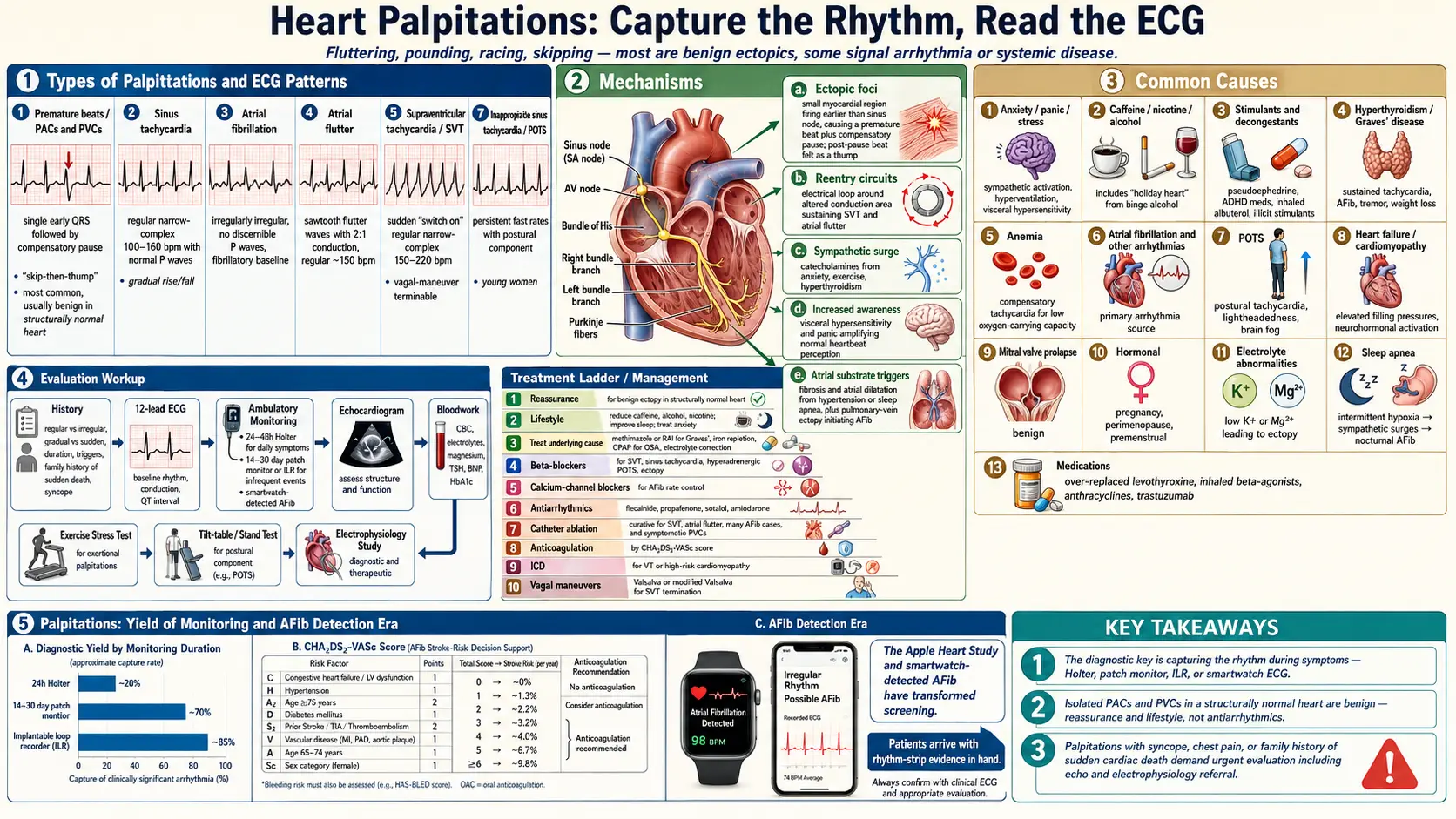

Table of Contents
- Overview
- Types of Palpitations
- Common Causes
- Arrhythmia Causes in Detail
- Non-Arrhythmia Cardiac Causes
- Metabolic and Endocrine Causes
- Pharmacological and Substance Causes
- Psychiatric and Functional Causes
- Mechanisms
- Evaluation
- Management
- Treatment by Cause
- When to Seek Medical Care
- Connections
- References & Research
- Featured Videos
Overview
Heart palpitations are the conscious awareness of the heart beating — described as fluttering, pounding, racing, skipping, or thumping. Most palpitations are benign and reflect ordinary ectopic beats (premature atrial or ventricular contractions), caffeine, sleep deprivation, anxiety, or hormonal shifts. A meaningful minority signal a treatable arrhythmia or systemic condition. The most useful question is what the palpitations do: a few skipped beats while resting feel different from a sudden rapid run lasting minutes, and the latter requires capture on a recording device for diagnosis.
Patients describe the sensation in their own vocabulary — "my heart was jumping out of my chest," "I felt a flip-flop," "it skipped a beat and then thudded," "my heart was racing for no reason." It is felt in the chest, throat, or neck; it may last a single beat or run for minutes to hours; and it can appear at rest, during exertion, or on lying down.
Palpitations are extremely common: they account for 1–3% of all primary care visits and are among the top five presenting complaints in cardiology clinics. Because they are almost always episodic, the resting ECG is frequently normal by the time the patient reaches the clinic — which is why the history and the choice of monitoring device carry most of the diagnostic weight.
Types of Palpitations
- Premature beats (PACs and PVCs) — the single most common cause; described as a skip, a hard beat, or a flip. Usually benign in the structurally normal heart.
- Sinus tachycardia — gradual rise and fall in rate, often tied to exertion, fever, anxiety, dehydration, or anemia.
- Atrial fibrillation — irregular and often rapid, may feel chaotic. Risk rises with age, hypertension, sleep apnea, alcohol, and hyperthyroidism.
- Atrial flutter — sudden onset, regular and rapid, often with 2:1 conduction.
- Supraventricular tachycardia (SVT) — sudden "switch on" of a regular fast rhythm at 150–220 bpm, often terminated by vagal maneuvers.
- Ventricular tachycardia — rapid, often associated with structural heart disease; medical emergency if sustained.
- Inappropriate sinus tachycardia and POTS — persistent fast rates with postural component, common in young women.
Common Causes
- Anxiety, panic, and stress — sympathetic activation, hyperventilation, and visceral hypersensitivity amplify ordinary heartbeat awareness.
- Caffeine, nicotine, alcohol — particularly binge alcohol intake ("holiday heart").
- Stimulants and decongestants — pseudoephedrine, ADHD medications, asthma inhalers (albuterol), illicit stimulants.
- Hyperthyroidism (Graves' disease) — produces sustained tachycardia, atrial fibrillation, tremor, and weight loss.
- Anemia — the heart compensates for low oxygen-carrying capacity by speeding up.
- Atrial fibrillation and other arrhythmias — structural and electrical disorders of the atria, AV node, ventricles.
- POTS — postural tachycardia, often with lightheadedness and brain fog.
- Heart failure and cardiomyopathy — can present with palpitations from arrhythmia or compensatory tachycardia.
- Mitral valve prolapse — benign, common, and a frequent source of palpitation awareness.
- Hormonal — pregnancy, perimenopause, premenstrual phase.
- Electrolyte abnormalities — low potassium or magnesium increases ectopy.
- Sleep apnea — intermittent hypoxia and surges in sympathetic tone trigger nocturnal palpitations and atrial fibrillation.
- Medications — thyroid replacement (over-replacement), inhaled beta-agonists, some chemotherapies (anthracyclines, trastuzumab).
- Pheochromocytoma, hypoglycemia, and fever — less common but readily identified once considered; each is described in the metabolic section below.
Arrhythmia Causes in Detail
True arrhythmias are the most clinically important cause of palpitations. The sensation produced varies by arrhythmia type, and the patient's description alone is sometimes enough to narrow the differential.
Premature Atrial Contractions (PACs)
PACs are the single most common cause of the "skipped beat" sensation. Paradoxically, the patient does not feel the PAC itself but the compensatory pause that follows it — the heart waits, then beats with extra force (post-extrasystolic potentiation), and that forceful beat is what registers as a "thump." PACs are visible on ECG as early P waves with altered morphology. They are essentially benign in the absence of structural heart disease. Common triggers include caffeine, alcohol, fatigue, stress, and nicotine. Reassurance is the primary intervention; beta-blockers can reduce frequency if symptoms are disabling.
Premature Ventricular Contractions (PVCs)
PVCs produce the same "skipped beat" or "flip-flop" sensation as PACs but arise from ventricular ectopic foci. They appear on ECG as wide, bizarre QRS complexes without a preceding P wave, followed by a compensatory pause. PVCs occurring in a regular alternating pattern with normal beats are called bigeminy; every third beat is trigeminy. PVCs are common in the general population and are benign when the PVC burden is low (under 10% of total beats) and the heart is structurally normal. However, frequent PVCs — particularly those exceeding 10–20% of total beats — can themselves cause a reversible PVC-induced cardiomyopathy with reduced ejection fraction. This makes quantifying PVC burden on a Holter monitor important in symptomatic patients.
Atrial Fibrillation (AFib)
AFib causes palpitations described as irregular, fast, and often sustained — "my heart was going completely haywire." On ECG it is characterized by absent P waves, an irregularly irregular ventricular response, and a fibrillatory baseline. AFib is the most common sustained cardiac arrhythmia, affecting roughly 6 million Americans. Beyond symptoms, it carries substantial stroke risk from atrial thrombus formation.
Supraventricular Tachycardia (SVT)
SVT presents as sudden-onset, sudden-offset rapid regular palpitations — often described as "the switch just flipped." Heart rates typically run 150–250 bpm. The most common mechanism is AV node re-entrant tachycardia (AVNRT), in which a re-entry circuit within the AV node causes rapid, regular conduction. SVT is generally not life-threatening in the absence of structural heart disease or WPW, but it can cause hemodynamic compromise at very high rates or with prolonged duration.
Wolff-Parkinson-White Syndrome (WPW)
WPW results from an accessory conduction pathway (the Bundle of Kent) that bypasses the AV node. On resting ECG, the signature findings are a short PR interval, a delta wave (slurred upstroke of the QRS), and a widened QRS. Most WPW patients experience SVT from re-entry involving the accessory pathway. The critical danger is atrial fibrillation in WPW: if AFib develops and conduction travels preferentially down the accessory pathway rather than the AV node, the rapid and irregular impulses can trigger ventricular fibrillation. This makes WPW with AFib a medical emergency. Critically, the standard AV node-blocking drugs — digoxin, verapamil, diltiazem, and adenosine are all contraindicated in AFib with WPW — because they preferentially block the AV node, leaving the accessory pathway unopposed and potentially accelerating conduction to dangerous rates. Treatment is IV procainamide or electrical cardioversion. Curative catheter ablation of the accessory pathway is the definitive treatment for symptomatic WPW.
Ventricular Tachycardia (VT)
VT is defined as three or more consecutive ventricular beats at a rate exceeding 100 bpm. On ECG it appears as a wide-complex tachycardia (QRS wider than 120 ms). VT in the setting of structural heart disease — post-infarction scar, dilated cardiomyopathy, hypertrophic cardiomyopathy — is potentially life-threatening and warrants aggressive evaluation, including consideration of an implantable cardioverter-defibrillator. Sustained VT can cause hemodynamic collapse and degenerate into ventricular fibrillation. Not every wide-complex tachycardia is VT (SVT with aberrant conduction is the main alternative), but in a patient with structural heart disease the safest assumption is always VT until proven otherwise.
Long QT Syndrome
Long QT syndrome (LQTS) — both congenital and acquired — predisposes to Torsades de Pointes, a distinctive polymorphic VT that can degenerate into ventricular fibrillation and sudden cardiac death. The QTc is prolonged when it exceeds 470 ms in women and 450 ms in men by the Bazett formula. Congenital LQTS involves mutations in cardiac ion channel genes (KCNQ1, KCNH2, SCN5A — LQT1, LQT2, LQT3). Acquired LQTS is commonly drug-induced, by antiarrhythmics, antipsychotics, antibiotics, and antihistamines. Hypokalemia and hypomagnesemia potentiate drug-induced QT prolongation.
Non-Arrhythmia Cardiac Causes
Sinus Tachycardia
Sinus tachycardia — a regular heart rate above 100 bpm driven by the sinus node — is the most common rapid heart rate overall. It is never a primary problem; it is always a physiological response to an underlying driver. The key clinical task is identifying the cause: pain, hypovolemia, sepsis, pulmonary embolism, heart failure, fever, anemia, hyperthyroidism, dehydration, anxiety, stimulants, or drugs. Sinus tachycardia should not be treated with rate-slowing agents in isolation — that suppresses a compensatory response without addressing the root problem. Patients typically describe a persistent "racing" feeling that builds gradually, rather than the sudden onset of SVT.
POTS (Postural Orthostatic Tachycardia Syndrome)
POTS is a form of dysautonomia defined by a heart rate increase of 30 bpm or more within 10 minutes of standing (or 40 bpm in adolescents), without orthostatic hypotension. Palpitations are a cardinal symptom — patients notice their heart racing whenever they stand up, often accompanied by lightheadedness, fatigue, brain fog, nausea, and near-syncope. POTS predominantly affects young women (female-to-male ratio approximately 5:1) and is increasingly recognized as a post-viral syndrome, including after COVID-19. Diagnosis is confirmed by an active stand test or a tilt table test.
Valvular Heart Disease
Mitral valve prolapse has historically been associated with palpitations; most prolapse is benign, and the palpitations are caused by concomitant PACs and PVCs rather than by the prolapse itself. Severe aortic regurgitation can cause prominent awareness of the heartbeat because of the large stroke volume and pounding pulse (Corrigan's pulse). Any significant valvular disease can also trigger atrial fibrillation, which then produces palpitations.
Metabolic and Endocrine Causes
Hyperthyroidism
Excess thyroid hormone sensitizes cardiac tissue to catecholamines and directly increases heart rate, stroke volume, and cardiac output. Patients experience fast, regular palpitations often present even at rest, along with heat intolerance, excessive sweating, unintended weight loss despite increased appetite, tremor, anxiety, diarrhea, and insomnia. TSH is suppressed and is the appropriate first-line screening test. Causes include Graves' disease (autoimmune, the most common), toxic multinodular goiter, and thyroiditis. Atrial fibrillation complicates hyperthyroidism in roughly 10–15% of cases and is a common initial presentation in older patients with "apathetic hyperthyroidism," who may lack the classic adrenergic features entirely.
Pheochromocytoma
Pheochromocytoma — a catecholamine-secreting tumor of the adrenal medulla — classically presents with episodic palpitations, severe headache, and diaphoresis (the classic triad), often accompanied by hypertensive crises. Episodes typically last minutes to an hour and may be triggered by positional change, physical exertion, or even abdominal palpation. Sustained hypertension can also occur. Screening uses plasma free metanephrines (sensitivity above 95%) or 24-hour urine fractionated metanephrines. CT or MRI of the adrenal glands follows a positive biochemical screen. Surgical resection is curative; alpha-blockade with phenoxybenzamine or doxazosin is essential preoperatively to prevent hypertensive crisis during induction of anesthesia.
Hypoglycemia
When blood glucose falls, the body mounts a sympathoadrenal counter-regulatory response — epinephrine and norepinephrine surge — producing palpitations, tremor, diaphoresis, and anxiety. Patients often wake at night feeling clammy and panicked. This is most relevant in people with diabetes using insulin or sulfonylureas, but it also occurs in reactive hypoglycemia after high-carbohydrate meals. Checking blood glucose during an episode, by fingerstick or continuous glucose monitor, is diagnostic. Treatment addresses the underlying cause: insulin dose adjustment, dietary change, or evaluation for rare endogenous hyperinsulinism (insulinoma).
Anemia
Anemia of any cause reduces oxygen-carrying capacity, and the compensatory rise in heart rate and stroke volume produces a hyperdynamic circulation with prominent palpitations, particularly on exertion. In severe anemia the heart "pounds" even at rest. A complete blood count is the key initial test; the underlying cause — iron deficiency, B12 or folate deficiency, hemolysis, chronic disease — guides treatment, and correcting the anemia resolves the palpitations.
Electrolyte Disturbances
Hypokalemia shifts the resting membrane potential and makes cardiac cells more excitable. PVCs are common; the ECG classically shows flattened T waves and prominent U waves. Hypokalemia is frequent in patients using loop diuretics (furosemide) or thiazides, in eating disorders with purging, and in diarrheal illness. Potassium replacement corrects most hypokalemia-associated palpitations.
Hypomagnesemia frequently co-occurs with hypokalemia and independently predisposes to arrhythmias, particularly Torsades de Pointes. Magnesium is essential for cardiac ion channel function and for the Na/K-ATPase pump that maintains intracellular potassium. IV magnesium is the first-line treatment for Torsades de Pointes regardless of the measured serum magnesium level. Common causes include diuretic use, alcoholism, malabsorption, and proton pump inhibitor use, which impairs intestinal magnesium absorption.
Fever
For every 1°C rise in core body temperature, heart rate increases by approximately 10 bpm. Any febrile illness can therefore cause prominent palpitations. The clinical task is identifying and treating the infection — the palpitations resolve with defervescence.
Pharmacological and Substance Causes
Caffeine
Caffeine is the most common dietary trigger. It blocks adenosine receptors, increasing sympathetic tone and directly exciting cardiac tissue, precipitating PACs and PVCs in susceptible individuals. The relationship is dose-dependent: moderate intake of up to 300–400 mg/day is generally tolerated, but individual sensitivity varies widely. Sources include coffee, tea, energy drinks, cola sodas, and dark chocolate.
Alcohol
Alcohol triggers palpitations through two distinct mechanisms. Acutely, even moderate intake can precipitate PACs and AFib in susceptible individuals. More dramatically, "Holiday Heart Syndrome" — first described by Ettinger in 1978 — refers to AFib or other atrial arrhythmias that develop after binge drinking in otherwise healthy young adults with no structural heart disease; the arrhythmia typically converts to sinus rhythm spontaneously as blood alcohol clears. Chronic heavy use causes alcoholic cardiomyopathy and its associated arrhythmias. Any patient with recurrent unexplained AFib should be asked carefully about alcohol intake.
Stimulant Medications
Multiple over-the-counter and prescription drugs contain sympathomimetic agents that raise heart rate:
- Decongestants — pseudoephedrine and phenylephrine in cold and allergy medications are alpha/beta agonists that raise heart rate and blood pressure; a commonly missed trigger.
- ADHD medications — amphetamine salts and methylphenidate are sympathomimetics; palpitations and elevated heart rate are documented side effects.
- Weight-loss pills — many contain caffeine, synephrine, or other stimulants; supplement labels need careful review.
Nicotine
Nicotine stimulates nicotinic acetylcholine receptors, acutely increasing heart rate and blood pressure through catecholamine release. Both cigarette smoking and nicotine replacement products — patch, gum, vaping — can trigger palpitations, particularly during a transition to higher-dose replacement.
Thyroid Hormone Over-Replacement
Patients on levothyroxine who are supraphysiologically dosed develop the palpitation pattern of hyperthyroidism: fast, regular, and present at rest. A TSH suppressed below 0.1 mIU/L on thyroid supplementation should prompt dose reduction. This is commonly seen after empiric dose increases without TSH follow-up.
Beta-Agonist Bronchodilators
Albuterol, salmeterol, and other beta-2 agonists used in asthma and COPD also stimulate cardiac beta-1 receptors — they are not perfectly selective — causing sinus tachycardia and palpitations. The effect is dose-dependent and worsens with overuse of rescue inhalers.
Antiarrhythmic Proarrhythmia
A paradoxical risk of antiarrhythmic drugs is that they can cause the very arrhythmias they are meant to treat. Class IC agents — flecainide and propafenone — are particularly notable: used for AFib, they can organize the fibrillation into atrial flutter that then conducts 1:1 to the ventricle at rates of 200–250 bpm. This is why class IC drugs for AFib are routinely combined with an AV node blocker. The CAST trial demonstrated that class IC agents increased mortality in post-infarction patients; they are contraindicated in structural heart disease.
Digoxin Toxicity
Digoxin has a narrow therapeutic window, with a target level of 0.5–0.9 ng/mL in heart failure. Toxicity — from accumulation, drug interactions with amiodarone or verapamil, or renal impairment — causes a wide range of arrhythmias: accelerated junctional rhythm, bidirectional VT (pathognomonic), and various degrees of AV block. Hypokalemia worsens digoxin toxicity. Management includes holding the drug, correcting electrolytes, and digoxin-immune Fab antibodies for severe toxicity.
Recreational Drugs
- Cocaine — blocks catecholamine reuptake, causing an intense sympathomimetic surge; associated with VT, AFib, coronary vasospasm, and myocardial infarction even in young patients without atherosclerosis.
- MDMA — releases massive serotonin, dopamine, and norepinephrine stores; causes tachycardia, hypertension, hyperthermia, and serious arrhythmias.
- Cannabis — acute use causes sinus tachycardia in most users; higher-potency products are increasingly associated with cannabis-associated myocardial infarction in young adults.
Psychiatric and Functional Causes
Panic Disorder and Panic Attacks
Panic disorder is a major cause of palpitations and one of the most important diagnoses to consider once cardiac causes have been excluded. A panic attack produces a sudden surge of autonomic activation — palpitations, shortness of breath, chest pain, dizziness, tingling, sweating, and an overwhelming sense of impending doom. Attacks peak at roughly 10 minutes and then subside. They can occur during the day or wake the patient from sleep as nocturnal panic attacks. Because the physical symptoms so closely mimic cardiac events, patients often present to emergency departments believing they are having a heart attack. An important diagnostic principle: panic disorder should be diagnosed only after arrhythmias have been adequately excluded — some patients have both a genuine arrhythmia and panic, and finding one does not rule out the other.
Generalized Anxiety Disorder
Patients with generalized anxiety disorder develop chronic autonomic hyperarousal — persistently elevated sympathetic tone producing resting tachycardia, palpitations, muscle tension, fatigue, and difficulty concentrating. Unlike the discrete episodes of panic, this causes more continuous background somatic symptoms, noticed particularly during periods of heightened worry, and somatic hypervigilance amplifies awareness of normal beats. Beta-blockers such as propranolol 10–20 mg as needed can interrupt the feedback loop in which noticing a palpitation causes anxiety, which causes more palpitations.
Somatic Symptom Disorder and Cardiac Anxiety
A subset of patients with palpitations have objectively normal Holter monitor findings — including during symptom episodes — with no correlation whatsoever between symptoms and rhythm. These patients often have high levels of health anxiety focused on the heart, amplified by hypervigilance to normal cardiac sensations. Central sensitization, in which the perception threshold for interoceptive signals is lowered, likely plays a role. Treatment focuses on cognitive behavioral therapy targeting cardiac anxiety, psychoeducation, and low-dose beta-blockers to reduce the peripheral somatic signals that feed the anxiety loop. Avoiding repeated cardiac testing, which reinforces illness beliefs, is part of the therapeutic approach.
Mechanisms
- Ectopic foci — a small region of myocardium fires earlier than the sinus node, producing a premature beat followed by a compensatory pause; the post-pause beat is often felt as a thump.
- Reentry circuits — an electrical impulse loops around an area of altered conduction, sustaining tachyarrhythmias such as SVT and atrial flutter.
- Sympathetic surge — catecholamines from anxiety, exercise, or hyperthyroidism increase rate and contractility.
- Increased awareness — visceral hypersensitivity and panic disorder amplify awareness of normal heartbeat.
- Triggers in atrial substrate — fibrosis, atrial dilatation (from hypertension or sleep apnea), and pulmonary-vein ectopy initiate atrial fibrillation.
Evaluation
- History — pattern (regular vs irregular), onset (gradual vs sudden), duration, triggers, family history of sudden death, syncope.
- 12-lead ECG — baseline rhythm, conduction abnormalities, QT interval.
- Ambulatory monitoring — 24- to 48-hour Holter for daily symptoms; 14- to 30-day patch monitor or implantable loop recorder for less frequent events.
- Echocardiogram — rule out structural heart disease, valvular disease, hypertrophy.
- Bloodwork — CBC, electrolytes, magnesium, TSH, BNP, HbA1c; fasting glucose when hypoglycemia is plausible, and plasma free metanephrines when pheochromocytoma is suspected.
- Exercise stress testing — if exertional palpitations or risk factors for ischemia; also detects exercise-induced VT and chronotropic incompetence.
- Tilt-table or stand test — if postural component; tracks heart rate and blood pressure response to passive head-up tilt.
- Electrophysiology study — for sustained or unexplained arrhythmias and to guide ablation, which can be performed in the same session.
Reading the Description
The character of the sensation maps onto the differential more reliably than any other single piece of information:
- Skipping or flip-flopping — PACs or PVCs.
- Racing and regular, switching on and off abruptly — SVT.
- Racing and irregular — atrial fibrillation.
- Pounding without a fast rate — heightened awareness of normal beats, as in anxiety, anemia, or aortic regurgitation.
- Gradual build and taper — sinus tachycardia; begins on standing — POTS.
- Associated symptoms — syncope or presyncope is the highest-risk companion; also ask about dyspnea, chest pain, diaphoresis, and neurological events suggesting embolic stroke from AFib.
- A complete medication list — including supplements, decongestants, and caffeine-containing products, which patients rarely volunteer.
What to Look For on the Resting ECG
A 12-lead ECG should be obtained in every patient presenting with palpitations, even if the episode has already resolved — several findings visible at rest fundamentally redirect the workup: WPW pre-excitation (delta wave with a PR interval under 120 ms), a prolonged QTc, ST-segment changes suggesting ischemia, pathological Q waves from prior infarction, a Brugada pattern (coved ST elevation in V1–V2), left ventricular hypertrophy suggesting hypertrophic cardiomyopathy, and PVCs present at rest.
Choosing an Ambulatory Monitor
The goal of ambulatory monitoring is a symptom-rhythm correlation — recording the rhythm during an episode to establish whether the patient's symptoms coincide with a documented arrhythmia or with a normal rhythm. Device choice follows episode frequency:
- Holter monitor — continuous recording, best for daily episodes; the patient keeps a symptom diary and analysis correlates the diary entries with rhythm strips.
- External event monitor or patch recorder — for less frequent episodes. Modern patch recorders record continuously for up to 14 days and combine algorithmic arrhythmia detection with patient event marking, giving better symptom-rhythm correlation than a Holter in many studies.
- Implantable loop recorder — a subcutaneous device placed under local anesthesia that monitors continuously for up to 3 years. Reserved for unexplained syncope or very infrequent severe episodes where shorter monitoring has been non-diagnostic; also used after cryptogenic stroke to detect paroxysmal AFib.
When to Order an Echocardiogram
Transthoracic echocardiography is indicated for palpitations accompanied by exertional symptoms or syncope, for any suggestion of structural heart disease on history, examination, or ECG, and whenever a sustained arrhythmia has been documented. It measures left ventricular ejection fraction, wall thickness (for hypertrophic cardiomyopathy), valvular function, and diastolic function.
Management
- Reassurance for benign ectopy — in a structurally normal heart with normal monitor and echo, isolated PACs and PVCs warrant lifestyle measures, not antiarrhythmics.
- Lifestyle changes — reduce caffeine, alcohol, nicotine, and stimulants; improve sleep; treat anxiety; manage weight.
- Treat the underlying condition — methimazole or radioactive iodine for Graves', iron repletion for anemia, CPAP for sleep apnea, electrolyte correction.
- Beta-blockers — rate control and symptom relief for SVT, sinus tachycardia, hyperadrenergic POTS, and benign ectopy.
- Calcium-channel blockers — rate control in atrial fibrillation when beta-blockers are not tolerated.
- Antiarrhythmic drugs — flecainide, propafenone, sotalol, amiodarone for rhythm control.
- Catheter ablation — curative for SVT, atrial flutter, and many cases of atrial fibrillation and symptomatic PVCs.
- Anticoagulation — for atrial fibrillation based on stroke risk (CHA2DS2-VASc).
- Implantable cardioverter-defibrillator — for ventricular tachycardia or high-risk cardiomyopathy.
- Vagal maneuvers — the Valsalva or modified Valsalva (lying back, leg raise) can terminate SVT.
Treatment by Cause
Supraventricular Tachycardia (AVNRT)
Acute termination proceeds stepwise. The modified Valsalva maneuver is the most effective vagal technique: the patient strains for 15 seconds, then the legs are passively elevated to 45° for 15 seconds while supine. The REVERT randomized trial found this approach roughly doubled the success rate of the standard Valsalva — 43% versus 17%. Ice-water facial immersion stimulates the diving reflex and is useful in children. Carotid sinus massage is an option but is contraindicated with a carotid bruit, recent stroke, or known carotid stenosis. If vagal maneuvers fail, IV adenosine 6 mg by rapid push with an immediate saline flush, repeated at 12 mg if the first dose fails, terminates most SVT by transiently blocking AV node conduction. For recurrent symptomatic SVT, catheter ablation of the re-entrant circuit achieves cure rates above 95% and is preferred over lifelong antiarrhythmic therapy in most guidelines.
Atrial Fibrillation
Three considerations run in parallel. Rate control aims for a resting ventricular rate below 110 bpm. Rhythm control restores and maintains sinus rhythm — flecainide or propafenone when there is no structural disease, amiodarone or dofetilide more broadly, or catheter ablation by pulmonary vein isolation; the EAST-AFNET 4 trial showed that early rhythm control reduces major cardiovascular events compared with rate control alone in newly diagnosed AFib. Stroke prevention follows the CHA2DS2-VASc score, and direct oral anticoagulants — rivaroxaban, apixaban, dabigatran, edoxaban — are preferred over warfarin in non-valvular AFib for their better safety profile.
Premature Ventricular Contractions
Low-burden PVCs in a structurally normal heart need only reassurance and trigger avoidance. For symptomatic PVCs not responding to conservative measures, beta-blockers reduce both frequency and symptoms. Once the burden passes the threshold described above — especially with a reduced ejection fraction — catheter ablation of the PVC focus can dramatically reduce burden and may reverse the associated cardiomyopathy.
Long QT Syndrome
Core management: avoid every QTc-prolonging medication (the CredibleMeds QTDrugs list is the authoritative reference and should be checked before prescribing any new drug); correct potassium and magnesium; use nadolol or propranolol specifically — not cardioselective beta-blockers — as first-line therapy for LQT1 and LQT2; avoid competitive sports in LQT1 and LQT2, where adrenergic surges are the trigger; and implant an ICD for patients with prior cardiac arrest, syncope despite beta-blockade, or high-risk genetic variants.
POTS
Non-pharmacological measures are foundational: aggressive salt loading of 8–10 g NaCl/day, fluid intake of 2–3 L/day, waist-high compression garments of at least 20–30 mmHg, and a structured exercise reconditioning program that begins with recumbent work — swimming or rowing — to avoid the orthostatic challenge of upright exercise. Drug options include fludrocortisone for volume expansion, low-dose propranolol at 10–20 mg for symptom relief, ivabradine off-label as a sinus-node If current inhibitor that lowers heart rate without impairing contractility or causing the fatigue common with beta-blockers, and midodrine, an alpha-1 agonist vasoconstrictor that reduces venous pooling.
Panic Disorder
First-line treatment combines an SSRI — sertraline, escitalopram, or paroxetine, the last carrying a higher discontinuation-syndrome risk — with cognitive behavioral therapy built around interoceptive exposure, in which palpitation-like sensations are deliberately induced to decondition the fear response. The combination achieves remission in 70–80% of patients. Short-acting benzodiazepines such as lorazepam 0.5–1 mg are appropriate as needed for acute severe attacks but should not anchor maintenance therapy, because of dependence risk. Beta-blockers help situational performance anxiety but are not effective maintenance treatment for panic disorder — they block the peripheral symptoms without touching the central fear circuitry.
Caffeine and Alcohol
A structured 2- to 4-week elimination trial of caffeine, alcohol, or both is simultaneously diagnostic and therapeutic: if the palpitations resolve with abstinence, the cause is identified. Where a trigger is confirmed, sustained avoidance is the most effective long-term intervention. For alcohol-associated AFib, randomized data show a substantial reduction in AFib burden and recurrence with abstinence in regular drinkers.
Metabolic Causes
Beta-blockers give rapid relief of the adrenergic symptoms of hyperthyroidism while definitive therapy takes effect, and palpitations plus AFib resolve on return to a euthyroid state. Potassium is repleted orally for mild to moderate hypokalemia and intravenously when severe or symptomatic; significant hypomagnesemia is repleted intravenously. Anemia is treated according to its etiology.
When to Seek Medical Care
- Palpitations with chest pain, pressure, or jaw or arm pain.
- Loss of consciousness or near-syncope during palpitations — the single most concerning companion symptom, raising the possibility of ventricular tachycardia, hypertrophic cardiomyopathy, or Brugada syndrome.
- Palpitations that last more than several minutes, or that recur frequently.
- Family history of sudden cardiac death, particularly under age 50 — which raises the possibility of an inherited channelopathy (long QT syndrome, Brugada syndrome, catecholaminergic polymorphic ventricular tachycardia) or hypertrophic cardiomyopathy.
- Palpitations that occur during exertion rather than after it — exercise-induced arrhythmias are more likely to be dangerous than resting ones and warrant stress testing.
- A prolonged QTc on any previous ECG — see the long QT section above for why this matters.
- Palpitations with new shortness of breath, leg swelling, or exercise intolerance.
- Known structural heart disease, prior heart attack, or cardiomyopathy — these sharply raise the probability that palpitations represent a ventricular arrhythmia.
- Palpitations with weight loss, tremor, and heat intolerance (rule out hyperthyroidism).
- Episodic palpitations with severe headache, drenching sweats, and high blood pressure (rule out pheochromocytoma).
Connections
- All Symptoms
- Anxiety
- Atrial Fibrillation
- POTS
- Cardiomyopathy
- Anemia
- Arrhythmia — the cardiology overview of the rhythm disturbances palpitations can signal.
- Heart Failure
- Heart Attack
- Obstructive Sleep Apnea
- Graves' Disease
- Hypertension
- Lightheadedness
- Dizziness
- Thyroid Disorders
- Magnesium
- Magnesium Deficiency and Heart Palpitations — how low magnesium makes the heart electrically irritable.
- Potassium
- Shortness of Breath
- Chest Pain
- Insomnia
- MSG
References & Research
Historical Background
Awareness of the heartbeat has been recorded since antiquity, but Holter's 1949 invention of the portable ECG monitor made systematic study of palpitations possible. The 21st century has produced increasingly sophisticated wearables, from patch monitors to consumer smartwatches that detect atrial fibrillation, transforming an occasionally elusive symptom into one frequently caught on tape before the doctor visit.
Key Research Papers
- Raviele A, Giada F, Bergfeldt L, et al. Management of patients with palpitations: a position paper from the European Heart Rhythm Association. Europace. 2011;13(7):920–934 — Search PubMed.
- Hindricks G, Potpara T, Dagres N, et al. 2020 ESC guidelines for the diagnosis and management of atrial fibrillation. European Heart Journal. 2021;42(5):373–498 — Search PubMed.
- Weber BE, Kapoor WN. Evaluation and outcomes of patients with palpitations. American Journal of Medicine. 1996;100(2):138–148 — Search PubMed.
- Page RL, Joglar JA, Caldwell MA, et al. 2015 ACC/AHA/HRS guideline for the management of adult patients with supraventricular tachycardia. Circulation. 2016;133(14):e506–e574 — Search PubMed.
- Perez MV, Mahaffey KW, Hedlin H, et al. Large-scale assessment of a smartwatch to identify atrial fibrillation. New England Journal of Medicine. 2019;381(20):1909–1917 — Search PubMed.
- January CT, Wann LS, Calkins H, et al. 2019 AHA/ACC/HRS focused update of the 2014 guideline for the management of patients with atrial fibrillation. Circulation. 2019;140(2):e125–e151 — Search PubMed.
- Marcus GM, Vittinghoff E, Whitman IR, et al. Acute consumption of alcohol and discrete atrial fibrillation events. Annals of Internal Medicine. 2021;174(11):1503–1509 — Search PubMed.
- Linz D, McEvoy RD, Cowie MR, et al. Sleep-disordered breathing and atrial fibrillation. JAMA Cardiology. 2018;3(6):532–540 — Search PubMed.
- Locati ET, Vecchi AM, Vargiu S, et al. Role of extended external loop recorders for the diagnosis of unexplained syncope, presyncope, and sustained palpitations. Pacing and Clinical Electrophysiology. 2014;37(12):1720–1728 — Search PubMed.
- Barsky AJ. Palpitations, arrhythmias, and awareness of cardiac activity. Annals of Internal Medicine. 2001;134(9 Pt 2):832–837 — Search PubMed.
- Giada F, Gulizia M, Francese M, et al. Recurrent unexplained palpitations (RUP) study: comparison of implantable loop recorder versus conventional diagnostic strategy. Journal of the American College of Cardiology. 2007;49(19):1951–1956 — Search PubMed.
- Appelboam A, Reuben A, Mann C, et al. Postural modification to the standard Valsalva manoeuvre for emergency treatment of supraventricular tachycardias (REVERT): a randomised controlled trial. Lancet. 2015;386(10005):1747–1753 — Search PubMed.
- Kirchhof P, Camm AJ, Goette A, et al. Early rhythm-control therapy in patients with atrial fibrillation (EAST-AFNET 4). New England Journal of Medicine. 2020;383(14):1305–1316 — Search PubMed.
- Blomström-Lundqvist C, Traykov V, Tabitha PA, et al. European Heart Rhythm Association (EHRA) consensus document on management of supraventricular arrhythmias. Europace. 2019;21(5):655–700 — Search PubMed.
- Gorenek BC, Buber J, Georgiou S, et al. Cardiac arrhythmias in acute coronary syndromes: position paper from the joint EHRA, ACCA, and EAPCI task force. Europace. 2014;16(11):1655–1673 — Search PubMed.
- Abbott AV. Diagnostic approach to palpitations. American Family Physician. 2005;71(4):743–750 — Search PubMed.
- Orejarena LA, Vidaillet H Jr, DeStefano F, et al. Paroxysmal supraventricular tachycardia in the general population. Journal of the American College of Cardiology. 1998;31(1):150–157 — Search PubMed.
- Brugada P, Brugada J, Mont L, Smeets J, Andries EW. A new approach to the differential diagnosis of a regular tachycardia with a wide QRS complex. Circulation. 1991;83(5):1649–1659 — Search PubMed.
- Maron BJ, McKenna WJ, Danielson GK, et al. American College of Cardiology/European Society of Cardiology clinical expert consensus document on hypertrophic cardiomyopathy. Journal of the American College of Cardiology. 2003;42(9):1687–1713 — Search PubMed.
- Priori SG, Napolitano C, Schwartz PJ. Low penetrance in the long-QT syndrome: clinical impact. Circulation. 1999;99(4):529–533 — Search PubMed.
- Grubb BP. Postural tachycardia syndrome. Circulation. 2008;117(21):2814–2817 — Search PubMed.
PubMed Topic Searches
- Evaluation of palpitations
- Atrial fibrillation screening with wearables
- Premature ventricular contractions: benign or pathologic?
- Hyperthyroidism and atrial fibrillation
- POTS and inappropriate sinus tachycardia
- Panic disorder and palpitations
- Pheochromocytoma, palpitations, and plasma metanephrines
- Wolff-Parkinson-White with atrial fibrillation: management